Dr Jean-Marc Chardonneau
A new treatment concept for new therapeutic actions.
Cellulite is the result of a ratio of power between dermal resistance and the pressure exerted inside the intra-lobular spaces in the adipose tissue, in the superficial hypodermis.
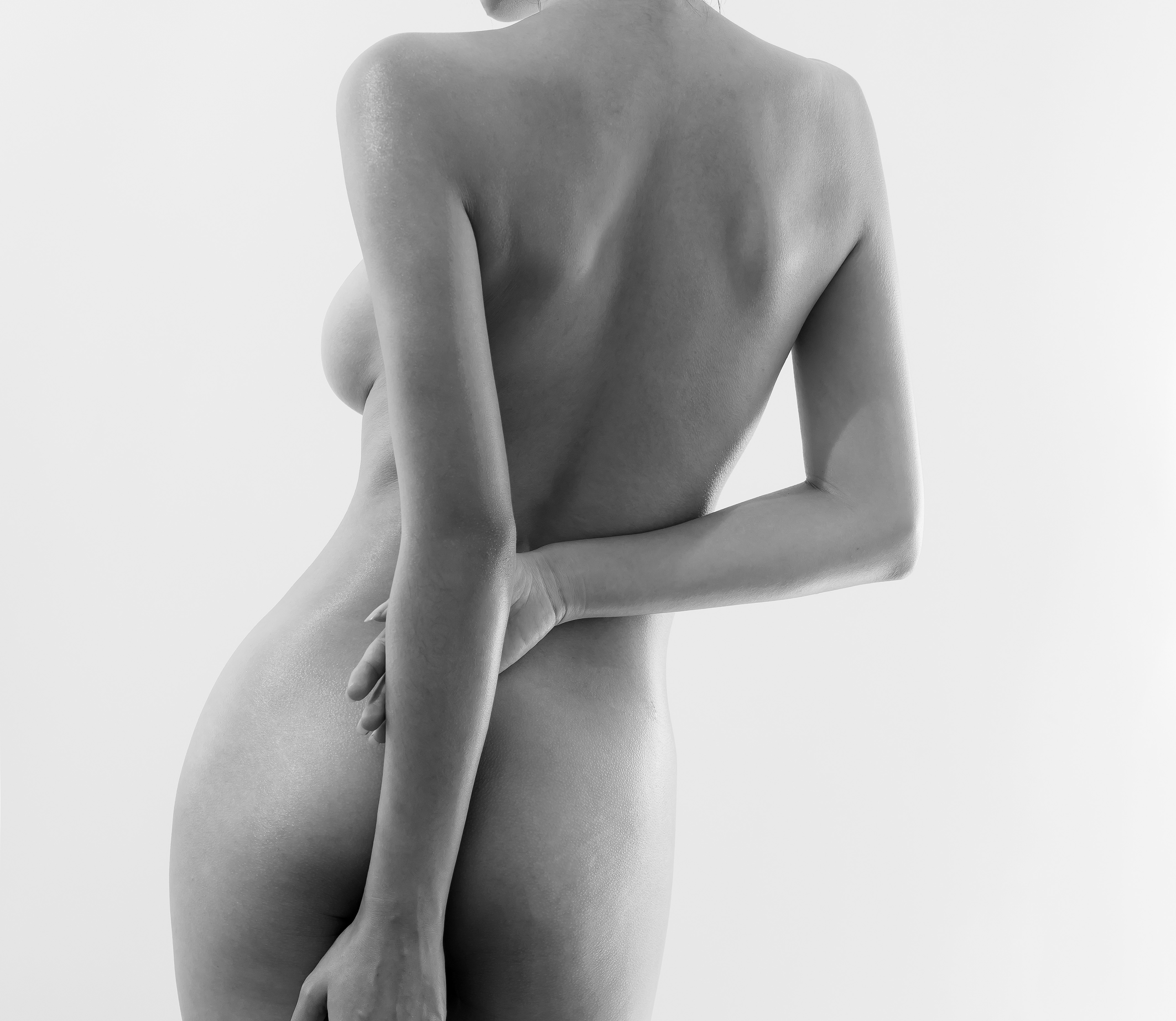 Schematically, underneath the dermis lies the hypodermis, the home of fatty tissue. The fat cells are grouped together in lobules. Between the lobules is the intra-lobular space through which the vessels, mainly lymphatic, and nerves run. The lymph – a water-like liquid that circulates in the intra-lobular spaces in the lymphatic vessels and towards the deeper layers – can meet resistance, which can prevent it from being evacuated and thus leads to this lymphatic liquid stagnating in the inter-lobular spaces. This dilation of the inter-lobular spaces explains the pressure exerted on the dermis and the clinical appearance of “orange-peel” skin.
These resistances or blockages can be split into 3 types: accumulation of fatty tissue (adipose cellulite), significant swelling (aqueous cellulite) and fibrosis (the hardening of the two types of cellulite previously mentioned). If there are no obstacles, it is the weakness of the dermis that has become very thin and lacks resistance (atonic cellulite). This is often the case for people who have been overexposed to the sun. Cellulite concerns two different entities
The diagnosis depends on the patient’s medical history, their clinical examination and, if possible, a scan. It must be said that the thing we call cellulite is actually two very different things. Real cellulite, with its orange-peel appearance, and steatoma, which is responsible for the curves that shape the body.
Both of these reside in the hypodermis. This little-known area is nothing more than a reservoir for fat.
It is divided into two parts that create a unit with the dermis (superficial hypodermis) and another adjacent to the mus- cular aponeurosis.
-
- With high-resolution scans (22 MHz), I recently showed that cellulite (orange-peel skin) is located in the superficial layer of the hypodermis, at a depth of 5 to 7mm
- Cellulite is a kind of oedematous-fibrous magma and is the result of an equation (see below) that opposes two forces: the resistance of the dermis on one side and the pressure exerted by the superficial hypodermis on the other. When the resistance of the dermis is less than the pressure exerted by the superficial hypodermis, cellulite develops.
The dermal resistance depends on the thickness of the dermis and the presence of normal-structured collagen fibres. The pressure exerted by the superficial hypodermis is linked to the pressure inside the walls that separate the fatty lobules. The presence of abnormal amounts of swelling and fibrosis in this space between the lobules is responsible for this pressure.
-
- The cause of this accumulation of swelling and fibrosis seems to be the microcirculation, namely too much permeability in the capillary vessels.
The pertinence of the diagnosis coupled with a suitable treatment is the key to success. A new therapeutic approach is completely in sync with the notions of physiopathology explained above: EndoSphere therapy or compressive micro-vibration. Developed by a team of Italian researchers and backed up by a number of studies, this therapy has indisputable potential for treating cellulite. The 55 spheres are positioned in a honeycomb pattern and rotate on themselves to produce micro-vibrations.
 Angiology graduate from the French National College of Aesthetic Medicine. DIU (interuniversity degree) in obesity, aging and aesthetics of superficial Tissue (Paris V). Graduated in Phlebology from the Paris V Marie Curie Institute and the French National College of Aesthetic Medicine (Paris VI). Inventor of the microsculpture and mesocannulation techniques for the legs. Consultant for Care, Solidea, Cutera servier, etc.