By Dr Diala Haykal
Chronological aging and photoaging: how to find the right solutions
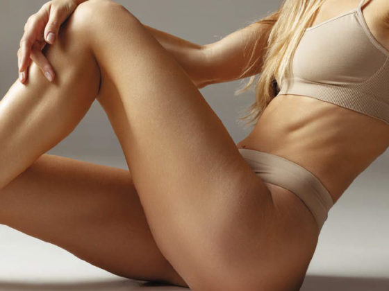
 Our skin, like all our other organs, suffers from chronological aging. In addition to this, unlike the other organs, the skin is in direct contact with the environment and is therefore affected by a type of aging caused by the sun’s UV rays.
Aging is a multifactorial process, determined both by our genes and by environmental factors which lead to anomalies in the genome and its expression (mutations, changes to the DNA’s repair capacity, shortening of the telomeres), alteration of the mitochondrial functions, oxidative stress (increased ROS – reactive oxygen species), alterations to the cell repair systems, etc.
Excess ROS are bad for the skin because they can cause oxidative damage in the cells, which leads to collagen breakdown. Ultraviolet A (320-400nm) is involved in the indirect production of ROS, leading to breaks in the DNA. Ultraviolet B (290320nm) leads to DNA mutations.
The clinical signs include wrinkles, lentigos, hyper-pigmentation, rosacea and reduced elasticity. These changes are more severe in people with a low phototype, and are influenced by their ethnic origin and their individual genes.
Photoaging can be avoided and addressed using a variety of treatments, including topical retinoids, chemical peels, injectables and energy-based treatments, or even a combination of the above.
Histological changes:
Photoaging causes a number of histological changes in the skin, which are different to the ones caused by chronological aging.
In chronologically-aged skin, the clusters of collagen fibres are short, fine and disorganised. The epidermis becomes thin, atrophic, and the dermo-epidermal junction is flattened. There are fewer melanocytes and Langerhans cells, in comparison with young skin that is protected from the sun. There is also a reduction in the length of the telomeres in the epidermis and dermis.
Photo-damaged skin is marked by increased elastosis, the fragmentation of the collagen underneath the dermo-epidermal junction. Its epidermal thickness can be irregular, there is an increased quantity of GAG glycosaminoglycans and proteoglycans, with an increased number of hyperplasic fibroblasts.
Unlike the hypocellular nature of chronologically-aged skin, photoaged skin can have an increased number of inflammatory cells such as mastocytes, eosinophils and mononuclear cells. The length of the telomeres is not influenced by extrinsic aging.
Clinical characteristics:
The skin, when aging chronologically, becomes thinner, with fine lines, xerosis, angiomas and seborrheic keratosis. Photoaged skin or heliodermia is atrophic with deep wrinkles, lentigos, a tanned appearance, telangiectasias, haematomas and dermatitis. The skin becomes thicker, with dilated follicular openings. Actinic keratoses and carcinomas may appear.
Therapeutic anti-aging strategies:
Anti-aging strategies have been developed with the aim of making the skin younger-looking and healthier. Applying sunscreen will protect the skin from the damaging effects of UV rays.
Medical research has led to the development of a diverse range of treatments that aim to rejuvenate the skin.
There are various anti-aging approaches:
Topical:
- Retinoic acid: Derivative of Vit A, promotes the proliferation of keratinocytes, strengthens the epidermis’ protective function, inhibits metalloprotein activity
- Ascorbic acid: reduces ROS, leads to collagen and elastin synthesis
- Glycolic acid: stimulates GAG and collagen production in the dermis
- Vit C: antioxidant, reduces ROS, neutralises oxidative stress
Energy-based devices
- Fractional lasers (ablative and non-ablative): stimulate the restructuring of the matrix
- Intense pulsed light (IPL): polychromatic pulsed light treats vascular and pigmented lesions, and improves neocollagenesis
- Light-emitting diodes (LED): treat skin inflammation and skin aging through photobiomodulation, trigger skin collagen synthesis and reduce metalloprotein expression
- Radiofrequency: provokes collagen contraction, skin firming, and improves the quality of the elastin fibres
- High-intensity focused ultrasounds (HIFU): lead to new collagen and elastin production in the deep reticular dermis
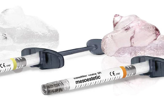
Injectables:
- Stimulate the fibroblasts, promote collagen synthesis, hydrate the skin
There has been a significant increase in the number of treatment options available for heliodermia in recent years.
For anyone who is looking for a miracle solution, avoiding sun exposure is the most effective treatment.
 Dr Diala HAYKAL, a private doctor established in Palaiseau since 2015, studied medicine at Paris VI university, where she completed a PhD in 2015, winning the Ageorges award for her thesis. Inter-university degree in Morphological and Anti-Aging Medicine from Paris Descartes University. University degree in Medical Lasers from Paris Descartes University. American Board of Aesthetic Medicine.Teacher and internship supervisor for the Paris VI universities, member of the Laser group of the French Society of Dermatology, member of the American Academy of Aesthetic Medicine. Founder of the Centre Médical Esthétique Laser Palaiseau.
More information esthetique-laser-palaiseau.fr/